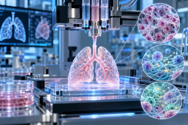
Researchers at the Indian Institute of Technology Hyderabad have developed a lung-derived bioink that can reproduce important biological and mechanical characteristics of natural lung tissue while supporting 3D bioprinting. The research introduces an osmotic decellularisation technique designed to preserve critical components of the lung extracellular matrix, creating a biologically active material that can guide stem-cell development and influence immune responses.
The study was conducted by Soham Ghosh and Professor Falguni Pati of the BioFab Lab in IIT Hyderabad’s Department of Biomedical Engineering. Published in the journal Biomaterials, the research addresses one of the central challenges in pulmonary tissue engineering: developing a printable material that is structurally stable while retaining the biochemical signals found in natural lung tissue.
Recreating the Lung’s Natural Cellular Environment
Human tissues contain an extracellular matrix, or ECM, consisting of proteins, carbohydrates and other biological molecules surrounding cells. Far from serving only as a structural framework, the extracellular matrix provides biochemical and mechanical signals that influence how cells grow, differentiate and behave.
This signalling role makes the ECM particularly valuable in regenerative medicine. Artificial materials can provide physical support for cells, but they often lack the complex biological cues present in natural tissue. Decellularised extracellular matrix materials attempt to overcome this limitation by removing cells from donated tissue while retaining as much of the underlying biological framework as possible.
For lung tissue engineering, however, the process presents an additional difficulty. Lung tissue is exceptionally soft, while a bioink used in extrusion-based 3D bioprinting must also be able to pass through a printer nozzle and regain sufficient structural stability to maintain the intended three-dimensional shape.
OsmoDcel Uses Osmotic Pressure Instead of Harsh Detergent Processing
The IIT Hyderabad researchers developed an osmotic decellularisation strategy called OsmoDcel. Instead of relying predominantly on conventional detergent-based methods, the technique subjects lung tissue to alternating hypo- and hypertonic conditions. Differences in salt concentration generate osmotic stress that helps rupture and remove cellular material.
The researchers designed the process to clear cells while preserving important components of the extracellular matrix. Their experiments showed retention of sulfated glycosaminoglycans, basement-membrane proteins, fibronectin and elastin networks.
This preservation is important because conventional detergent-based decellularisation can remove useful extracellular components along with unwanted cellular material. Losing these molecules can alter the mechanical properties of the resulting material and reduce biological signals capable of influencing cell behaviour.
The researchers compared their osmotic method with modified conventional detergent-based lung decellularisation techniques, including processes involving sodium dodecyl sulphate and Triton X-100 with sodium deoxycholate.
Turning Decellularised Lung Matrix Into a Printable Bioink
After decellularisation, the preserved lung extracellular matrix was processed into a material suitable for extrusion-based 3D bioprinting. The team studied how the bioink behaved while flowing through a printer nozzle and how rapidly its internal structure recovered once the mechanical force was removed.
The researchers used print-matched thixotropy testing rather than relying solely on conventional bulk-viscosity measurements. This allowed them to examine conditions closer to those experienced by the bioink during actual printing, including nozzle residence time and wall shear stress.
Their experiments revealed different advantages depending on how much of the original lung matrix composition was preserved. Collagen-dominant formulations demonstrated stronger vertical layer stacking because of their higher viscosity, while matrices retaining larger amounts of sulfated glycosaminoglycans rebuilt their viscosity more rapidly and gelled quickly after extrusion.
The resulting hydrogels remained within mechanical ranges relevant to the soft lung parenchyma while maintaining compatibility with living cells during the bioprinting process.
Bioink Guides Stem Cells Towards Lung-Relevant Cell Types
One of the most significant findings concerned the behaviour of mesenchymal stem cells placed within or on the bioprinted lung matrix.
The researchers found that cells responded differently depending on their position within the printed scaffold. Stem cells seeded on the surface developed markers associated with epithelial differentiation, including cytokeratin and tight-junction proteins, together with transcripts associated with pulmonary surfactant.
Cells encapsulated deeper within the matrix developed expression of alpha-smooth muscle actin, or α-SMA, consistent with an interstitial myogenic phenotype.
These results indicate that the decellularised lung extracellular matrix does more than provide a physical structure. It retains biological information capable of influencing cell fate, an important property for researchers attempting to reproduce the complex organisation of living lung tissue.
Encouraging an Anti-Inflammatory Immune Environment
The IIT Hyderabad team also examined interactions between the bioprinted material and macrophages, immune cells that play an important role in inflammation, tissue repair and regeneration.
The OsmoDcel-based constructs promoted a macrophage response associated with a pro-regenerative and anti-inflammatory environment, including when the researchers subjected the system to an inflammatory challenge in laboratory experiments.
This immunomodulatory behaviour is important for regenerative medicine because an implanted or engineered tissue construct must interact successfully with the body’s immune system. Excessive or persistent inflammation can interfere with healing and compromise tissue regeneration.
By combining cellular guidance with favourable immune responses, the lung-derived bioink provides a platform for investigating how engineered tissue materials can influence both regeneration and inflammation.
Applications in Lung Tissue Engineering and Disease Research
The technology remains an experimental research platform rather than a clinical treatment. However, the work has potential applications across regenerative medicine, pulmonary disease modelling, drug testing and fundamental studies of lung biology.
Bioprinted lung-like tissue can provide researchers with laboratory models that reproduce aspects of human tissue architecture more realistically than conventional two-dimensional cell cultures. Such systems could eventually improve studies of respiratory diseases and provide more physiologically relevant platforms for evaluating drug candidates.
The approach may also contribute to the broader development of patient-specific tissue models and regenerative strategies in which biological materials actively influence cellular development rather than merely supporting cell growth.
IIT Hyderabad Strengthens Its Biofabrication Research Programme
The work forms part of IIT Hyderabad’s growing research programme in biofabrication and tissue engineering. The institute’s Department of Biomedical Engineering lists 3D bioprinting, tissue and organ models, novel bioink development, regenerative medicine and personalised medical technologies among the principal research areas of its Biofabrication and Tissue Engineering group.
IIT Hyderabad has also established an Indo-German Bioengineering Centre of Excellence focused on lung disease research. The collaboration brings together IIT Hyderabad and Germany’s Institute for Lung Health with the objective of developing bioengineered lung and vascular models and advancing personalised approaches to pulmonary disease.
The OsmoDcel research adds an important materials platform to this broader effort. By demonstrating that careful preservation of the natural lung extracellular matrix can produce a printable material capable of directing stem-cell behaviour while supporting a regenerative immune environment, the study strengthens India’s growing capabilities in 3D bioprinting, biomaterials and regenerative medicine.
References
Indian Institute of Technology Hyderabad, Department of Biomedical Engineering — Biofabrication and Tissue Engineering Research
https://bme.iith.ac.in/assets/pages/rese.html
Indian Institute of Technology Hyderabad — ILH-IITH Bioengineering Centre of Excellence
https://iith.ac.in/events/2026/01/07/bioengineering-coe-inauguration/
Ghosh S, Pati F. “Bioprinted cell-instructive lung-dECM constructs for reconstructing an immuno-regenerative pulmonary microenvironment.” Biomaterials. DOI: 10.1016/j.biomaterials.2026.124508
https://pubmed.ncbi.nlm.nih.gov/42561800/
Elsevier, Biomaterials — Original Research Article
https://www.sciencedirect.com/science/article/pii/S0142961226005326
You may also like
-
Indian Scientists Discover Molecular Switch That Can Regulate Cellular Autophagy
-
Scientists Map Earliest Glow of Hydrogen, Opening A New Window to the Universe
-
Indian Researchers Study RAD51 DNA-Repair Gene Variant in Prostate Cancer
-
TDB Backs Multi Nano Sense With ₹37.51 Crore to Build Indigenous Multi-Gas Sensor Platform
-
IISER Berhampur and NIT Rourkela Researchers Explore Cotarnine Derivatives for Wound Healing and Biomedical Applications
