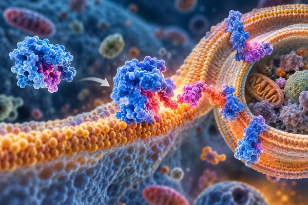
Indian researchers have identified a previously unknown molecular mechanism that can switch the activity of LC3, a key protein involved in autophagy, the cellular process responsible for capturing and recycling damaged proteins, organelles and other unwanted material. The discovery provides scientists with a new way to experimentally control an important stage of the cell’s internal recycling machinery and could support future research into diseases in which autophagy becomes disrupted.
The research was led by scientists from the CSIR-Institute of Genomics and Integrative Biology in New Delhi, working with researchers from the Academy of Scientific and Innovative Research, National Institute of Immunology, Ashoka University and the University of California, Los Angeles. The peer-reviewed study, titled A programmable lipid-triggered allosteric site modulates LC3 LIR receptor binding activity, was published in Nature Communications on August 28, 2026.
Autophagy Acts as the Cell’s Internal Recycling System
Autophagy is a fundamental biological process through which cells remove damaged or unnecessary components. Material identified for removal is enclosed inside specialised double-membrane structures called autophagosomes. These structures subsequently deliver their contents for degradation and recycling, allowing useful molecular components to be reused.
The process is essential for maintaining normal cellular function. Problems in autophagy have been associated with several areas of biomedical research, including cancer, neurodegenerative disorders, metabolic diseases, infections and ageing. Understanding precisely how the process is regulated therefore remains an important field of cell biology.
A protein known as LC3 plays a central role in autophagy. Once attached to the autophagosome membrane, LC3 acts as a docking platform for receptor proteins that recognise cellular material destined for removal. These receptors contain LC3-interacting regions, commonly known as LIR motifs, which connect selected cargo with the developing autophagosome.
Scientists already understood many aspects of how LC3 interacts with these receptors. What remained less clear was how attachment to the autophagosome membrane itself changes the protein and regulates its ability to capture cargo.
Membrane Contact Changes the Shape of LC3
The research team found that LC3 undergoes a substantial structural change when it interacts with a lipid membrane. Before reaching the membrane, important functional pockets within the protein remain relatively inaccessible. Membrane attachment causes LC3 to rearrange its structure, exposing regions required for interaction with autophagy receptors.
This means that membrane attachment does not simply hold LC3 in the correct location. It also acts as a molecular signal that changes the protein into a more functionally active state.
The researchers traced this transformation to a previously identified structural region within LC3 that behaves as an allosteric site. Allostery occurs when a change at one part of a protein alters the behaviour of another region located some distance away. Such mechanisms allow proteins to operate like molecular switches, with structural changes transmitting signals across the molecule.
In LC3, the newly characterised allosteric mechanism connects membrane interaction with the receptor-binding regions responsible for recognising autophagy cargo.
Scientists Engineer Active and Inactive Versions of LC3
To determine whether the newly identified allosteric site could actually control LC3 activity, the researchers used molecular-dynamics simulations and protein-design techniques to modify the region.
From several engineered variants, the team identified two particularly important forms of LC3. One mutation stabilised the membrane-bound protein in an active configuration, while another stabilised it in an inactive configuration.
The active variant displayed stronger receptor binding and promoted the capture of cellular cargo. The inactive version remained attached to the membrane but showed greatly reduced functional activity.
This distinction demonstrated that the researchers could alter LC3’s cargo-recruitment behaviour without simply preventing it from reaching the autophagosome membrane. Instead, the protein’s activity could be controlled through its internal structural switch.
Multiple Techniques Confirm the Molecular Mechanism
The researchers combined computational and experimental methods to verify the mechanism. Molecular-dynamics simulations were used to examine the movement of LC3 and identify structural communication between the allosteric region and its functional binding pockets.
The engineered protein variants were then investigated using X-ray crystallography, which enabled researchers to examine their structures at atomic resolution. Biophysical experiments were used to measure protein behaviour and receptor interactions.
Super-resolution microscopy provided further evidence of the way LC3 behaved within cellular environments, while transmission electron microscopy helped examine its relationship with membrane structures and cargo capture.
Together, these approaches confirmed that activation of the allosteric region encourages receptor binding and cargo recruitment, while stabilising the inactive state substantially reduces these functions.
A Programmable Molecular Switch for Autophagy
One of the most important aspects of the discovery is the possibility of making LC3 activity programmable. By altering a relatively small structural region of the protein, researchers demonstrated that the autophagy machinery could be shifted towards either an active or inactive state.
The work therefore provides a new experimental tool for investigating selective autophagy, in which cells identify and remove particular proteins, aggregates, organelles or other material rather than indiscriminately degrading cellular components.
Selective autophagy relies heavily on the interaction between LC3 and specialised cargo receptors. The newly discovered allosteric mechanism offers researchers a way to examine these interactions while independently controlling the functional state of LC3.
CSIR Files Patent Application for the LC3 Molecular Switch
The research has also produced intellectual property related to the engineered LC3 system. According to the published study, the Council of Scientific and Industrial Research is the applicant for a patent covering the design of a mutant LC3B allosteric switch for regulating autophagy.
Researchers Lipi Thukral, Deepanshi Gahlot and Niyati Jain are listed as inventors on the patent application. The international application, PCT/IN2026/050284, was reported as pending when the research paper was published.
The patent activity highlights the potential value of the molecular switch as a research platform for controlling and studying autophagy rather than merely observing the process after it occurs.
CSIR-IGIB Research Combines Computational Biology and Experimental Science
The project reflects the interdisciplinary approach of the CSIR-Institute of Genomics and Integrative Biology. Lipi Thukral, a scientist at CSIR-IGIB and professor at AcSIR, leads research involving computational structural biology, molecular dynamics and the study of interactions between proteins and lipids.
Her research programme examines how molecular motion influences the structure and function of biomolecules, particularly proteins associated with cellular membranes. High-performance computing and molecular-dynamics simulations are combined with experimental techniques to understand protein behaviour at extremely small spatial and temporal scales.
The LC3 study extends this work by showing that membrane-associated proteins can undergo structural changes that fundamentally alter their biological activity.
Discovery Could Open New Directions in Disease Research
The findings do not constitute a treatment for cancer, Alzheimer’s disease or other conditions associated with disrupted autophagy. The research is instead a fundamental advance in understanding how one of the cell’s major recycling proteins is regulated.
Its importance lies in providing researchers with a mechanism that can be deliberately manipulated. Scientists studying diseases involving defective protein clearance, abnormal organelle accumulation or altered autophagy could use such molecular switches to investigate how increasing or reducing particular stages of the pathway affects cellular behaviour.
The principle may also extend beyond LC3. Many proteins interact with cellular membranes, and membrane contact can alter their shape and activity. Understanding these membrane-triggered structural mechanisms could therefore reveal similar regulatory switches in other biological pathways.
By showing that a lipid membrane can activate a hidden structural switch within LC3, and that the switch itself can be engineered to regulate receptor binding and cargo capture, the CSIR-IGIB-led team has provided a new molecular framework for studying selective autophagy. The work strengthens India’s growing capabilities in computational structural biology, protein engineering and fundamental biomedical research while opening new avenues for understanding one of the cell’s most important maintenance systems.
References
CSIR-Institute of Genomics and Integrative Biology — Lipi Thukral, Computational Structural Biology
https://www.igib.res.in/Scientists/lipi-thukral
Nature Communications — A programmable lipid-triggered allosteric site modulates LC3 LIR receptor binding activity
https://www.nature.com/articles/s41467-026-76697-9
PubMed — Gahlot D, Castin J, Mathur S, et al. A programmable lipid-triggered allosteric site modulates LC3 LIR receptor binding activity
https://pubmed.ncbi.nlm.nih.gov/42665581/
DOI
https://doi.org/10.1038/s41467-026-76697-9
You may also like
-
Scientists Map Earliest Glow of Hydrogen, Opening A New Window to the Universe
-
Indian Researchers Study RAD51 DNA-Repair Gene Variant in Prostate Cancer
-
TDB Backs Multi Nano Sense With ₹37.51 Crore to Build Indigenous Multi-Gas Sensor Platform
-
IISER Berhampur and NIT Rourkela Researchers Explore Cotarnine Derivatives for Wound Healing and Biomedical Applications
-
IISER Tirupati Develops pH-Responsive DNA Nanostructures for Controlled Drug Delivery
