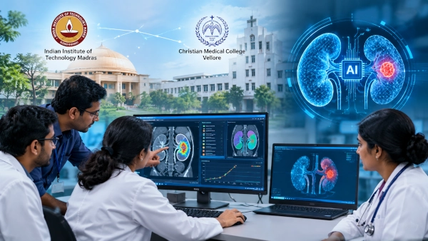
Researchers at the Indian Institute of Technology Madras and Christian Medical College, Vellore, have developed a suite of artificial-intelligence tools designed to help doctors identify kidney disease earlier, classify abnormalities in medical scans and assess the extent of tumours with greater precision.
The collaboration combines engineering and machine-learning expertise from IIT Madras with clinical knowledge from CMC Vellore and addresses one of the most difficult problems in kidney care: many kidney disorders can progress for a considerable period before producing obvious symptoms.
By the time patients develop clear signs of serious kidney dysfunction, substantial damage may already have occurred. Earlier identification can therefore give clinicians more time to investigate the underlying cause, monitor disease progression and begin appropriate treatment before irreversible loss of kidney function develops.
The research team has developed three complementary AI-based technologies, each addressing a different stage of kidney assessment.
AI Model Predicts Chronic Kidney Disease Risk
The first system uses machine learning together with clinical and laboratory information to estimate a patient’s risk of chronic kidney disease, or CKD.
Rather than depending on a single measurement, the approach allows multiple pieces of clinical information to be analysed together by an algorithm. Such systems could eventually help clinicians identify patients whose combination of laboratory values and health indicators suggests a higher probability of kidney disease.
The researchers have also developed a prototype interface intended to make the system easier to use in a clinical environment.
This type of predictive technology could become especially useful for screening patients who already have major CKD risk factors, including diabetes and hypertension. However, the system is intended to assist clinicians rather than replace established blood tests, urine investigations or medical judgement.
The larger objective is to provide doctors with a rapid and consistent decision-support tool capable of highlighting patients who may require closer examination.
Deep Learning Analyses More Than 12,000 Kidney CT Images
The second technology uses deep learning to analyse CT scans automatically.
The system has been trained on more than 12,000 kidney images and can classify scans into four categories: a normal kidney, kidney cyst, kidney stone or kidney tumour.
This is an important distinction because abnormalities visible in kidney imaging can have very different clinical implications. Kidney stones, cysts and tumours require different forms of investigation and treatment, while normal scans should ideally be recognised without unnecessary intervention.
A deep-learning system can be trained to recognise complex patterns in images by examining large numbers of examples. Once sufficiently validated, such technology could assist radiologists and other clinicians by rapidly screening scans and highlighting abnormalities that deserve closer attention.
The technology could be particularly useful in healthcare environments where large imaging volumes place pressure on specialists.
However, the research should not yet be interpreted as an autonomous diagnostic replacement for radiologists. The team plans further clinical validation across multiple centres and datasets before wider translation into routine healthcare.
3D Platform Measures Kidney Tumour Burden
The third component moves beyond simple image classification.
Researchers have developed an open-source 3D anatomical imaging platform capable of reconstructing a patient’s kidney from CT data. The resulting three-dimensional model can be used to calculate tumour volume and estimate how much of the kidney is affected by disease.
Conventional medical images are generally viewed as a series of two-dimensional slices. Experienced clinicians can interpret those images, but a three-dimensional reconstruction can provide a more intuitive representation of the kidney and the location and extent of a tumour.
This could be valuable when doctors are trying to understand the burden of disease or plan treatment.
Tumour size alone does not always provide a complete picture. Knowing how a tumour is distributed through the organ and what proportion of kidney tissue is involved could potentially provide additional patient-specific information for clinical decision-making.
Because the platform is based on open-source technology, it may also offer a relatively accessible route for further research and development compared with highly specialised proprietary imaging systems.
Bringing Engineering and Clinical Medicine Together
The project is being led by Prof. G.L. Samuel and research scholar Jennifer Delighta from IIT Madras, working with Prof. Santosh Varughese of CMC Vellore.
The collaboration is significant because medical AI systems require more than sophisticated algorithms. Engineers may be able to build highly accurate computational models, but those systems must also answer clinically useful questions and work with the realities of hospital data.
By combining engineering, artificial intelligence, imaging and nephrology expertise, the project attempts to develop tools around actual medical requirements.
The researchers say the objective is to give clinicians rapid and standardised diagnostic support, enabling them to make better-informed decisions while potentially allowing intervention at an earlier stage of disease.
Why Earlier Kidney Disease Detection Matters
Kidneys perform several essential functions, including filtering waste from the blood, regulating fluid and electrolyte balance and contributing to the control of blood pressure.
When kidney function declines gradually, the body can initially compensate. This means chronic kidney disease may remain largely unnoticed during its earlier stages.
Advanced disease can eventually require expensive and demanding interventions such as dialysis, while some patients may require transplantation.
Technology that helps identify high-risk patients earlier could therefore have value both for individual patients and for the wider healthcare system.
The IIT Madras-CMC Vellore project approaches the problem from several directions rather than attempting to create one universal diagnostic algorithm.
The CKD risk model examines clinical and laboratory information. The CT classifier identifies major structural abnormalities, while the 3D imaging system provides a more detailed assessment of tumour involvement.
Together, the three systems could eventually form different layers of an integrated digital kidney-assessment platform.
Towards a Digital Twin of the Kidney
The longer-term ambition is particularly interesting.
The researchers describe the current work as a step towards developing a kidney “Digital Twin” — a digital representation of an individual patient’s organ that could combine medical imaging, clinical information and potentially continuous physiological data.
A sophisticated medical digital twin could theoretically be updated as new patient information becomes available, allowing clinicians to track disease progression and explore how an individual patient’s condition is changing over time.
The current project has not yet reached that stage. The three technologies are building blocks towards the concept rather than a completed digital twin system.
Nevertheless, the direction shows how medical artificial intelligence is moving beyond simple image recognition towards patient-specific computational models.
The work has been supported by IIT Madras and the Scheme for Promotion of Academic and Research Collaboration, or SPARC.
Wearables Could Add Continuous Health Data
The next stage of research will involve broader clinical validation using multi-centre datasets.
This is essential because an AI model that performs well using data from one institution may not necessarily perform equally well when exposed to patients, scanners and clinical practices from different hospitals.
Testing across multiple centres can help researchers determine whether the algorithms remain reliable across a more diverse population.
The IIT Madras-CMC team also plans to explore integration with wearable sensing platforms for long-term health monitoring.
If successful, such integration could eventually allow periodic hospital data to be combined with continuously collected physiological information. That would move kidney monitoring away from isolated clinical snapshots towards a more longitudinal view of a patient’s health.
AI as a Clinical Assistant, Not a Replacement
The most realistic near-term role for these systems is as clinical decision-support technology.
AI can examine large volumes of information quickly, detect patterns and provide standardised measurements. Doctors, however, must interpret those findings alongside a patient’s symptoms, history, laboratory investigations and other medical factors.
That distinction is especially important in healthcare, where a technically accurate algorithm may still require substantial validation before it can safely influence treatment.
The IIT Madras and CMC Vellore programme therefore represents an encouraging research development rather than a finished hospital diagnostic product.
Its significance lies in integrating three complementary capabilities — risk prediction, automated CT interpretation and patient-specific 3D modelling — within a single kidney-disease research programme.
If the technologies continue to perform successfully through multi-centre clinical validation, they could ultimately provide doctors with faster and more detailed information while helping identify kidney disease before extensive damage occurs.
More broadly, the research demonstrates how India’s engineering institutes and major clinical centres can work together to translate artificial intelligence from laboratory algorithms into tools designed around real medical problems.
The eventual kidney Digital Twin envisioned by the team remains a longer-term objective, but the three technologies developed so far provide an important foundation: identifying risk, recognising disease and measuring precisely how much of the organ has been affected.
You may also like
-
Why Ayurveda Warns Against Suppressing Hunger, Thirst and Sleep
-
India Strengthens Quality Standards for Ayurveda, Yoga and Sowa-Rigpa Through Nationwide BIS Initiative
-
How Indigenous Indian Technologies Are Helping Aircraft and Trains Navigate Safely Through Dense Fog
-
Sanofi Receives Indian Approval for Qfitlia, a New RNA-Based Treatment for Haemophilia A and B
-
Dr. Reddy’s Receives Indian Approval for Zytorvi Immunotherapy to Treat Advanced Oesophageal Cancer
