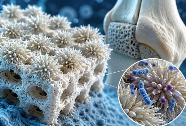
Indian researchers have turned to an unlikely source of inspiration in the search for safer bone implants: the spiny geometry of a sea urchin. A team associated with the Indian Institute of Technology Mandi (IIT Mandi) has developed a nano-structured hydroxyapatite coating for 3D-printed bone scaffolds that can physically damage bacteria while simultaneously creating a surface better suited for bone integration.
The research represents an interesting convergence of 3D printing, biomaterials, nanostructured surfaces and regenerative medicine. Instead of adding an antibiotic or conventional antibacterial chemical to the implant, the researchers engineered the physical structure of its surface so that microscopic spikes attack bacteria mechanically.
The study, titled “Nano-urchin ceramic coating: Topography mediated infection control and osteointegration in 3D-printed scaffolds,” was published online on July 17, 2026, in the Chemical Engineering Journal. The research was conducted by Ankita Negi, Aakash Verma, K. M. Mohammed Sufiyan, Vedante Mishra and Sumit Murab. IIT Mandi’s Translational Tissue Engineering Laboratory lists the paper among its latest publications.
The Two Problems Facing Bone Implants
Repairing large bone defects remains difficult because an implanted structure has to perform several functions at once. It must provide sufficient structural support, interact safely with surrounding tissue and gradually establish a strong interface with newly forming bone.
At the same time, an implanted material creates a potential surface for bacterial colonisation. An infection around an implant can complicate healing and, in serious cases, contribute to failure of the implant. The researchers therefore focused on two interconnected challenges: preventing bacterial infection and encouraging osteointegration, or the successful integration of an implant with bone.
Three-dimensional printing offers an attractive route for producing scaffolds shaped for particular bone defects. The IIT Mandi researchers used polylactic acid, or PLA, a thermoplastic material that can be 3D printed. However, untreated thermoplastic scaffolds can have relatively hydrophobic and biologically inactive surfaces, limiting their interaction with cells and tissue.
The team’s solution was not simply to change the shape of the scaffold. Instead, it engineered the surface at a much smaller scale.
Building a Sea Urchin-Like Surface
The researchers functionalised the 3D-printed PLA scaffold with hydroxyapatite, commonly abbreviated as HA. Hydroxyapatite is particularly relevant to bone engineering because it is closely related to the mineral component of natural bone.
The process begins with an alkali treatment of the PLA surface. This creates negatively charged chemical sites that can act as nucleation points for subsequent mineral formation. The researchers describe this process as resembling the way collagen provides a template for mineral deposition during natural bone formation.
A low-temperature hydrothermal process then allows biomimetic hydroxyapatite to precipitate directly on the scaffold.
Crucially, the hydroxyapatite does not form as a simple smooth layer. It develops into clusters containing numerous extremely small projections or nanospicules. Viewed together, these structures resemble the spikes extending from a sea urchin.
This unusual architecture gives the coating high porosity, increased surface area and considerable surface roughness. It also transforms the scaffold into a superhydrophilic surface, improving its interaction with water and the surrounding biological environment.
Using Geometry to Attack Bacteria
The most striking aspect of the research is how the coating attacks bacteria.
Many antibacterial implant strategies rely on antibiotics, antibacterial drugs, metallic ions or other chemical agents. The IIT Mandi approach instead exploits mechanobactericidal action.
When bacteria encounter the sea urchin-like coating, the nanospicules interact physically with their cellular structure. The geometry of the surface can damage bacterial cells without requiring the coating to release a conventional antibiotic. The research therefore explores a fundamentally different route to creating an infection-resistant implant surface.
The researchers tested the coated HA-PLA scaffolds against two important bacterial species: Escherichia coli and Staphylococcus aureus.
According to the reported results, the HA-PLA scaffolds achieved approximately 90% bactericidal activity against E. coli and 50% against S. aureus.
The team observed mechanobactericidal effects both in laboratory experiments and in an in-vivo subcutaneous mouse infection model, providing evidence that the physical antibacterial effect was not confined solely to a laboratory culture plate.
That distinction is important. The technology is not simply making it more difficult for bacteria to attach to an implant. Its engineered topography is intended to create a surface capable of physically damaging bacterial cells.
Helping Bone Grow at the Same Time
An antibacterial surface alone would not solve the implant problem. Bone must also be able to interact successfully with the scaffold.
This is where hydroxyapatite performs the second part of the technology’s function.
The researchers tested the scaffolds using MG-63 cells, a cell line widely used in studies of bone-related biological behaviour. The experiments indicated that the HA-coated scaffolds supported increased deposition of collagen and calcium, both important indicators associated with the development of a bone-supporting environment.
The team also investigated the material through subcutaneous ectopic implantation in mice. The results again showed enhanced collagen and calcium deposition on the HA-PLA scaffolds, supporting the researchers’ conclusion that the surface has the potential to promote bone formation.
The coating therefore performs two very different tasks through the same engineered architecture. Its nanospicules create an antibacterial physical surface, while the hydroxyapatite provides a mineral interface favourable to bone integration.
That combination is what makes the work particularly noteworthy.
Moving Beyond Antibiotic-Loaded Implant Coatings
Antibiotics remain essential medicines for treating bacterial infections. However, implant-associated infections present particular difficulties because bacteria can establish persistent colonies on implanted surfaces.
The IIT Mandi research explores whether the physical design of the material itself can form part of the antibacterial defence.
Rather than treating an implant as an inert object that subsequently needs antibacterial drugs added to it, the researchers are effectively engineering antibacterial behaviour into its microscopic architecture.
In this context, “antibiotic-free” refers specifically to the mechanism of the experimental coating. The sea urchin-like hydroxyapatite structure does not depend on incorporating an antibiotic into the scaffold to produce its reported mechanobactericidal effect. It should not be interpreted as evidence that antibiotics would no longer be required for patients who develop implant-associated infections.
3D Printing Adds Another Important Dimension
The underlying scaffold is produced using 3D printing, which adds another potentially important advantage.
Large bone defects vary considerably between patients. Additive manufacturing can potentially create scaffolds tailored to the geometry of a particular defect rather than forcing surgeons to work exclusively with standardised shapes.
The surface engineering technique could therefore complement the customisation offered by 3D printing. The broader concept is a patient-specific printed scaffold whose microscopic surface is independently engineered for biological integration and bacterial resistance.
This is precisely the type of intersection between advanced manufacturing and biomedical science that could eventually make additive manufacturing more useful in regenerative medicine.
IIT Mandi’s Translational Tissue Engineering Laboratory itself focuses on the interface between materials science and medicine, including biomaterial systems for 3D printing and bioprinting, injectable materials for orthopaedic tissue regeneration and biosimilar material platforms.
Research Built Over Several Years
The nano-urchin work also appears to be part of a longer research programme rather than an isolated experiment.
IIT Mandi’s publication record shows earlier work by the group on osteogenic interfaces for 3D-printed thermoplastic scaffolds, antibacterial coatings, acellular mineralisation and biomaterials for bone regeneration. The laboratory also records earlier presentations specifically investigating self-assembled hydroxyapatite nano-spicule coatings on 3D-printed PLA scaffolds.
An earlier version of the sea urchin concept was presented at the Bio-MANTHAN 2025 conference as a “Mechanobactericidal Osteogenic Sea Urchin-like Hydroxyapatite Nanospicule Coating on 3D Printed PLA scaffolds,” where it received a Best Oral Presentation Award.
The publication in the Chemical Engineering Journal therefore represents the progression of a research direction that the IIT Mandi team has been developing through successive studies.
Promising, But Still Preclinical
The results are encouraging, but the distinction between a successful experimental biomaterial and a clinically available implant is important.
The published work includes cell-culture experiments and mouse models. It does not establish that sea urchin-coated PLA implants are ready for routine implantation in human patients. Further research would be required to evaluate long-term safety, mechanical performance, degradation, infection control, bone regeneration and effectiveness in clinically relevant bone-defect models before eventual human trials could be considered.
Nevertheless, the research demonstrates an elegant principle: the surface of an implant need not remain passive.
By controlling architecture at extremely small scales, researchers can potentially give a material additional biological functions without relying solely on drugs or chemical antibacterial agents.
Nature’s Geometry Meets Indian Biomedical Engineering
The sea urchin-inspired coating illustrates how biomimicry can produce practical engineering ideas. Nature has evolved complex structures across scales, and scientists increasingly study such geometries for solutions to problems ranging from adhesion and fluid movement to structural strength and microbial control.
At IIT Mandi, that principle has been applied to one of medicine’s persistent challenges.
A 3D-printed polymer provides the basic scaffold. Hydroxyapatite creates a bone-compatible mineral surface. Nano-scale spikes inspired by the appearance of a sea urchin add a physical antibacterial mechanism.
The result is an experimental implant platform designed not merely to occupy the space left by damaged bone, but to actively shape the biological environment around it.
References
IIT Mandi – Translational Tissue Engineering Laboratory, Murab Group: The official IIT Mandi laboratory page identifies Dr Sumit Murab as Associate Professor in the School of Biosciences & Bioengineering and lists the nano-urchin research among the laboratory’s latest work.
IIT Mandi – Murab Group Publications: Official laboratory publication record listing Ankita Negi, Aakash Verma, K. M. Mohammed Sufiyan, Vedante Mishra and Sumit Murab as authors of the study published in the Chemical Engineering Journal on July 17, 2026.
Chemical Engineering Journal / Elsevier: Nano-urchin ceramic coating: Topography mediated infection control and osteointegration in 3D-printed scaffolds, Article 179600, DOI 10.1016/j.cej.2026.179600.
You may also like
-
ISRO Completes Historic SPADEX Mission as Both Satellites Re-enter Earth’s Atmosphere After Successful Space Docking Experiments
-
Indian Scientists Discover Rare Accretion Disk Around Neutron Star 24,000 Light-Years Away
-
BARC Scientists Develop Indigenous Technology to Recover 90% of Lithium from Seawater
-
How Indigenous Indian Technologies Are Helping Aircraft and Trains Navigate Safely Through Dense Fog
-
ISRO Successfully Completes Critical Gaganyaan Crew Module Separation Tests at VSSC
