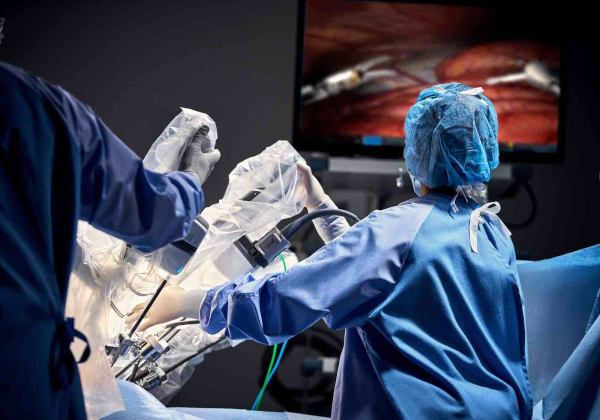
A new partnership between medical technology company Stryker and the Indian Association of Gynaecological Endoscopists (IAGE) will seek to expand surgeon training in fluorescence-guided surgery across India, bringing greater attention to an imaging technology that is increasingly being used to help surgeons visualise lymphatic pathways, blood flow and critical anatomical structures during minimally invasive gynaecological procedures.
The collaboration was announced on 31 August 2026 after an agreement was signed during the Pelvic Organ Prolapse Conference 2026 in Chennai. Around 350 gynaecological surgeons and healthcare professionals attended the conference. Under the initiative, Stryker and IAGE plan to conduct clinical education programmes across India over the next two years, including training and interaction with Indian and international specialists experienced in fluorescence-guided surgery.
The programme is significant because fluorescence-guided surgery, or FGS, represents an important evolution in minimally invasive surgery. Conventional laparoscopy provides surgeons with a magnified colour image of the operative field. Fluorescence imaging adds another layer of information by allowing selected tissues, blood vessels or lymphatic pathways to become visible under specialised near-infrared imaging.
Rather than replacing a surgeon’s anatomical knowledge or conventional imaging, fluorescence functions as a real-time navigation aid during an operation.
What Is Fluorescence-Guided Surgery?
Fluorescence-guided surgery combines a fluorescent imaging agent with a specialised camera capable of detecting wavelengths of light that cannot normally be seen by the human eye.
One of the most widely used agents is indocyanine green, or ICG, a fluorescent dye with a long history of medical use. Once administered, ICG can be illuminated using near-infrared light. The dye absorbs this energy and emits fluorescence at another near-infrared wavelength, which is captured by the surgical imaging system and displayed on a monitor.
The surgeon can therefore switch between normal visible-light images and fluorescence images during an operation.
Depending on where and how ICG is administered, the technique can provide different kinds of information. Intravenous ICG can highlight blood vessels and tissue perfusion, while carefully placed local injections can trace lymphatic drainage and reveal sentinel lymph nodes.
The U.S. Food and Drug Administration, for example, recognises ICG fluorescence imaging for visualisation of vessels, blood flow and tissue perfusion and specifically provides dosing instructions for lymphatic mapping in cervical and uterine cancer.
This ability to reveal information that is difficult to appreciate with ordinary white-light imaging explains why fluorescence technology is attracting increasing interest across surgery.
Sentinel Lymph-Node Mapping: One of Its Most Important Gynaecological Uses
One of the most established applications of fluorescence-guided surgery in gynaecology is sentinel lymph-node mapping in gynaecological cancers.
Cancer cells can spread from a primary tumour through the lymphatic system. Traditionally, determining whether this spread has occurred may require removal of multiple pelvic lymph nodes.
A sentinel lymph node is the first lymph node, or one of the first nodes, into which lymphatic fluid from a tumour-bearing organ is likely to drain. If this node can be accurately located and examined, surgeons may be able to obtain important staging information while avoiding a more extensive lymph-node dissection in appropriately selected patients.
With ICG-guided mapping, small quantities of the fluorescent agent can be injected into the cervix. The dye travels through lymphatic channels, which become visible under near-infrared imaging and can lead the surgeon towards the sentinel nodes.
This application is no longer merely experimental.
The 2025 ESGO-ESTRO-ESP guidelines for endometrial carcinoma recommend sentinel lymph-node biopsy for staging in patients with presumed uterus-confined disease and state that indocyanine green with cervical injection is the preferred detection technique.
Fluorescence can consequently help transform a difficult anatomical search into a visually guided procedure.
Instead of relying exclusively on normal tissue appearance, the surgeon sees a fluorescent lymphatic pathway leading towards the node that needs to be removed and examined.
Potential to Reduce the Extent of Surgery
The broader objective behind sentinel-node surgery is not simply better imaging. It is to obtain the staging information needed to guide cancer treatment while potentially avoiding unnecessary removal of large numbers of lymph nodes.
Extensive lymphadenectomy can be associated with complications including lymphatic fluid collections and lower-limb lymphoedema. Sentinel-node approaches are designed to reduce surgical morbidity when oncologically appropriate while preserving accurate staging.
Fluorescence imaging is particularly valuable here because successful sentinel-node surgery depends heavily on finding the correct nodes.
Research and expert consensus have found ICG to be a reliable lymphatic tracer in endometrial and cervical cancer, with particularly strong performance in bilateral sentinel-node detection.
The fluorescence system therefore does not treat cancer itself. Its role is to give the surgeon better visual information during the operation, potentially allowing surgery to become more targeted.
Seeing Blood Flow in Real Time
Another important capability of ICG fluorescence is assessment of tissue perfusion.
When ICG is administered intravenously, it circulates through the bloodstream. A near-infrared camera can then display how quickly and effectively blood reaches a particular piece of tissue.
This can be useful because tissue that appears acceptable under ordinary white light may nevertheless have compromised blood supply.
The FDA recognises ICG fluorescence imaging for visualisation of vessels, blood flow and tissue perfusion with an appropriately authorised imaging system. Fluorescence in blood vessels can become visible within seconds following intravenous administration.
In gynaecological surgery, perfusion assessment has potential applications in reconstructive surgery, difficult hysterectomies and operations involving the bowel.
It may be especially valuable during surgery for deep endometriosis, where disease can involve the rectum or other portions of the bowel and sometimes requires removal and reconstruction of affected tissue.
After a bowel segment has been reconnected, surgeons need confidence that the remaining tissue has an adequate blood supply. Fluorescence angiography can provide additional information about perfusion before the operation is completed.
Studies have found the technique promising for intraoperative bowel assessment in gynaecological and endometriosis surgery, although larger controlled studies are still required to establish how much it reduces complications in routine clinical practice.
Making the Ureters Easier to Identify
A particularly interesting emerging use of fluorescence imaging is ureter identification.
The ureters are narrow tubes carrying urine from the kidneys to the bladder. They run through areas of the pelvis in which gynaecologists routinely operate.
During straightforward surgery, their anatomical course can usually be identified conventionally. But operations involving severe endometriosis, extensive adhesions, previous surgery, large pelvic masses or altered anatomy can make identification considerably more difficult.
Accidental ureteric injury is uncommon but potentially serious.
Researchers have consequently investigated whether fluorescence could make the ureters more visible throughout difficult pelvic surgery.
A systematic review published in 2026 examined intraureteral ICG fluorescence for ureter identification during gynaecological procedures. Seven studies involving 299 patients met its criteria. All reported successful ureter visualisation, with no ureteral injuries or ICG-related complications reported in the included studies.
However, the authors stressed that the evidence remains limited. Most available studies are small and observational, and certainty regarding outcomes such as reductions in injury, blood loss or operating time remains low.
Ureter fluorescence should therefore be described as a promising developing application rather than a universally established standard of care.
It is also technically distinct from the established cervical injection of ICG used for lymphatic mapping in uterine and cervical cancers.
Particularly Interesting for Deep Endometriosis
Fluorescence-guided surgery may eventually have an important role in some of the most complicated cases of deep endometriosis.
Deep endometriosis can distort normal pelvic anatomy and affect structures including the bowel, ureters, bladder and tissues around the uterus. The challenge for surgeons is not simply removing disease. They must remove as much clinically significant disease as appropriate while preserving healthy organs, nerves and blood supply.
Fluorescence imaging has consequently been investigated for several purposes during endometriosis surgery: identifying ureters, assessing bowel perfusion, helping with anatomical orientation and, experimentally, improving visualisation of endometriotic lesions.
A 2026 systematic review of intraoperative navigation in endometriosis surgery found that ICG-based imaging is among the most commonly reported navigation technologies and can assist with anatomical orientation and perfusion-related decisions during difficult procedures. At the same time, the review concluded that the clinical evidence remains limited and that prospective comparative trials are needed to establish exactly where the technology provides the greatest benefit.
That distinction matters.
Fluorescence is already well established for some applications, particularly sentinel lymph-node mapping. Its usefulness for finding or defining endometriosis itself remains much less settled and should not be presented as though fluorescent imaging can reliably illuminate every endometriotic lesion.
Fluorescence Does Not Make Everything Glow
The phrase fluorescence-guided surgery can give the misleading impression that a camera automatically distinguishes diseased tissue from healthy tissue.
That is not how most current ICG-based systems work.
ICG is not generally a tumour-specific agent. Depending on how it is administered, it principally reveals physiological features such as blood flow or lymphatic drainage.
The surgeon must interpret that information together with normal visual imaging, preoperative scans, anatomy, tactile or instrument feedback and clinical judgement.
A fluorescent lymphatic channel can help locate a sentinel node. Fluorescent blood flow can indicate that tissue is perfused. Fluorescent marking of a ureter can make its course easier to follow.
But the image does not independently decide which tissue should be removed.
FGS is therefore better understood as augmented surgical vision rather than automated surgery.
Why Near-Infrared Imaging Is Useful
Near-infrared light has characteristics that make it useful for surgery.
Visible light is heavily affected by colour and surface tissue. Near-infrared wavelengths can travel somewhat deeper through biological tissue and are less affected by some forms of visible-light interference.
Special cameras can detect the fluorescence even though the surgeon cannot see the emitted near-infrared light directly.
Modern laparoscopic imaging platforms combine high-resolution conventional imaging with fluorescence modes, enabling surgeons to alternate rapidly between the ordinary operative view and fluorescence-enhanced information.
Stryker itself markets surgical visualisation platforms incorporating fluorescence capability, including its 1688 AIM 4K platform and newer 1788 minimally invasive surgery platform. Its Indian product information says the latter incorporates fluorescence imaging intended to improve visualisation of perfusion and critical anatomy.
The Stryker-IAGE agreement, however, is primarily an education and training partnership; the announcement should not be interpreted as evidence that a particular imaging platform is becoming mandatory or standard across Indian hospitals.
Training Is Critical to Using the Technology Properly
This is where the new IAGE partnership becomes important.
Fluorescence-guided surgery involves more than installing a new camera in an operating theatre. Surgeons must understand when to administer a fluorescent agent, where to administer it, how long to wait before imaging, how to interpret fluorescence patterns and when the fluorescent signal may be misleading.
Technique can differ substantially according to the clinical objective.
Sentinel-node mapping requires a different approach from intravenous perfusion imaging. Ureter identification uses still another technique. Each requires appropriate patient selection and an understanding of the evidence supporting the procedure.
Image quality can also be influenced by the amount of dye used, injection technique, timing, tissue characteristics, distance between the camera and tissue and the performance of the imaging platform.
Standardised training therefore becomes especially important as fluorescence moves beyond highly specialised centres into wider clinical practice.
Under the new partnership, Stryker and IAGE plan to conduct pan-India clinical education initiatives over the next two years, with participating surgeons receiving exposure to Indian and international specialists and established surgical practices.
From Minimally Invasive Surgery to Image-Guided Surgery
Laparoscopic and robotic surgery have already changed gynaecology by allowing complex operations to be conducted through small incisions with cameras providing magnified views inside the abdomen and pelvis.
Fluorescence represents another stage of that evolution.
A conventional laparoscopic camera primarily shows what a structure looks like. Fluorescence can add information about where lymph is draining, whether blood is reaching tissue or where a difficult anatomical structure lies.
That distinction is important because some of the greatest challenges in surgery involve information that ordinary vision alone cannot provide.
In cancer surgery, fluorescence can guide a surgeon towards the lymph node most relevant for staging. During reconstructive surgery, it can provide additional evidence about whether tissue has adequate perfusion. During difficult pelvic dissection, it may make important structures easier to identify.
The ultimate objective is not simply to create a brighter or more technologically impressive image. It is to allow the surgeon to make a better-informed decision while the operation is actually taking place.
Evidence Varies Between Applications
The growing enthusiasm around fluorescence-guided surgery should nevertheless be balanced against the quality of evidence.
Sentinel lymph-node mapping with ICG in endometrial cancer has become well established and is incorporated into major clinical guidelines.
Other applications remain at different stages of development.
Fluorescence assessment of tissue perfusion is established as an imaging capability, but the degree to which its use improves particular gynaecological outcomes can differ between operations. Fluorescent ureter identification is promising, particularly for complex pelvic surgery, but current systematic reviews still call for stronger prospective evidence. Using fluorescence to identify endometriotic disease itself remains an evolving research field.
This makes education particularly important. Surgeons need to understand not only what fluorescence imaging can do, but equally where evidence does not yet justify assuming a clinical benefit.
Building Capability Across Indian Gynaecological Surgery
The Stryker-IAGE collaboration should therefore be viewed principally as a surgeon-capability and clinical-education initiative.
IAGE is a professional organisation focused on gynaecological endoscopy and minimally invasive surgery, while Stryker develops surgical imaging and other medical technologies. Their two-year programme seeks to create structured opportunities for Indian surgeons to develop familiarity with fluorescence-guided techniques and exchange experience with specialists already using the technology.
If programmes of this kind succeed in broadening access to properly structured training, they could help fluorescence-guided techniques move beyond a relatively small number of high-end surgical centres.
That could be particularly relevant as minimally invasive gynaecological oncology and complex endometriosis surgery expand across India.
Yet the value of the technology will ultimately depend not on the number of hospitals possessing fluorescence-capable cameras, but on appropriate indications, trained surgeons, standardised technique and evidence-based use.
Fluorescence-guided surgery is not a replacement for surgical expertise. Its promise lies in giving that expertise additional information at precisely the moment when difficult decisions must be made.
For gynaecology, its clearest impact can already be seen in sentinel lymph-node mapping for cancer. Its expanding use for perfusion assessment, ureter identification and complex pelvic surgery suggests that operating theatres are gradually moving from an era of minimally invasive surgery towards a broader model of real-time image-guided precision surgery.
You may also like
-
Jangli Badam (Terminalia catappa): Indian Almond in Traditional Medicine, Nutrition and Tropical Healing
-
Udumbara — Ayurveda’s Cluster Fig for Wounds, Bleeding and Pitta-Kapha Balance
-
India Cuts Malaria Cases and Deaths by Nearly 80% as 160 Districts Report Zero Indigenous Transmission
-
Karanja — Ayurveda’s Bitter Oil Tree for Skin, Wounds and Kapha-Vata Balance
-
Bengaluru’s SugarStrings.ai Develops Cheek-Swab DNA Test Screening Thousands of Genetic Disorders
