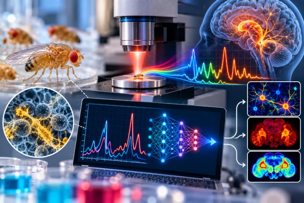
Indian researchers have demonstrated a rapid, label-free method for identifying biochemical changes associated with Parkinson’s disease by combining infrared spectroscopy with machine learning.
The study was carried out by researchers from the CSIR-Central Institute of Medicinal and Aromatic Plants in Lucknow and the Academy of Scientific and Innovative Research. The work used Drosophila melanogaster, the common fruit fly, as a laboratory model of Parkinson’s-like neurodegeneration.
The research has been published in Spectrochimica Acta Part A: Molecular and Biomolecular Spectroscopy and was highlighted by Research Matters on October 5, 2026.
Researchers Created Three Parkinson’s-Like Fruit Fly Models
The team induced Parkinson’s-like changes in fruit flies using three established neurotoxicants: paraquat, rotenone and MPTP.
These compounds are commonly used in experimental models because they can trigger features associated with Parkinson’s pathology, including oxidative stress, mitochondrial dysfunction and degeneration of dopamine-producing neurons.
The researchers first confirmed that the treated flies had developed relevant biological and behavioural changes before testing the infrared method. The flies showed impaired movement, disturbances in activity patterns and degeneration of dopaminergic neurons.
Infrared Spectroscopy Created a Biochemical Fingerprint
The core technology used in the study was attenuated total reflectance Fourier transform infrared spectroscopy, or ATR-FTIR.
Infrared spectroscopy works by measuring how biological molecules absorb infrared light at different wavelengths. Proteins, lipids, carbohydrates, nucleic acids and other cellular components absorb energy in characteristic patterns, producing a spectrum that acts like a biochemical fingerprint.
Instead of measuring one molecule at a time, FTIR provides a broader view of the overall molecular composition of a sample.
The researchers analysed intact fruit fly heads across the mid-infrared spectrum and found clear differences between healthy control flies and the Parkinson’s-like models.
Parkinson’s Models Showed Changes in Lipids and Proteins
The infrared spectra revealed alterations in regions associated with lipids, proteins and other cellular biomolecules.
Among the most important changes were signals linked with lipid metabolism and oxidative stress. The study identified notable spectral variations in the biological fingerprint region between 1012 and 1040 cm⁻¹, as well as changes in lipid-associated and protein-related regions.
These spectral changes were consistent with other evidence showing mitochondrial dysfunction, increased reactive oxygen species, lipid accumulation and neurodegeneration in the toxin-exposed flies.
Machine Learning Separated Healthy and Parkinson’s-Like Flies
Infrared spectroscopy can generate large datasets containing many overlapping signals, making interpretation difficult through conventional visual analysis alone.
The researchers therefore applied machine-learning and chemometric techniques to identify patterns within the spectra.
Methods included principal component analysis, partial least squares discriminant analysis, random forest and k-nearest-neighbour classification. These approaches were able to separate control samples from Parkinson’s-like models using biochemical features detected by infrared spectroscopy.
Importantly, the researchers also tested the models on independent datasets that had not been used during training, helping assess whether the classification approach could generalise beyond the original samples.
The Method Does Not Detect a Single Parkinson’s Biomarker
The study should not be interpreted as the discovery of a single diagnostic biomarker for Parkinson’s disease.
Instead, ATR-FTIR captures a combined biochemical signature reflecting changes across multiple molecular classes.
This is important because Parkinson’s disease involves many interacting biological processes, including oxidative stress, mitochondrial damage, altered lipid metabolism and the progressive loss of dopaminergic neurons.
The infrared approach therefore provides a broad biochemical snapshot rather than identifying one disease-specific molecule.
Why Fruit Flies Are Used in Parkinson’s Research
Drosophila melanogaster is widely used in neurodegeneration research because many important cellular pathways are conserved between fruit flies and humans.
Their short lifespan, well-characterised genetics and measurable behavioural responses also allow researchers to study disease mechanisms quickly and under controlled conditions.
Fruit fly Parkinson’s models cannot reproduce the full complexity of the human disease, but they provide an efficient experimental system for testing biological mechanisms and screening potential diagnostic or therapeutic approaches.
Potential for Faster Neurodegeneration Screening
Conventional biochemical studies often require targeted assays designed to measure specific molecules.
ATR-FTIR offers a different approach. A sample can be rapidly scanned without fluorescent labels or extensive chemical processing, producing a broad spectral profile within a relatively short period.
When combined with machine learning, subtle differences that may be difficult to recognise manually can be detected automatically.
This makes the method potentially useful for high-throughput neurotoxicity studies, drug screening and laboratory research into neurodegenerative disease.
Early Diagnosis Remains a Major Challenge in Parkinson’s Disease
Parkinson’s disease develops progressively, and cellular changes can begin long before obvious movement symptoms appear.
By the time characteristic symptoms such as tremor, rigidity and slowed movement become clinically apparent, substantial neurological damage may already have occurred.
Researchers worldwide are therefore searching for biomarkers that could reveal the disease earlier or help monitor its progression.
Spectral fingerprinting could eventually contribute to that effort by identifying biochemical changes associated with neurodegeneration before major clinical symptoms appear.
Human Clinical Use Is Still a Future Step
The current study was performed in chemically induced fruit fly models, not in people with Parkinson’s disease.
That distinction is important. A technique capable of distinguishing experimental models does not automatically become a clinical diagnostic test.
Future studies will need to evaluate larger and more diverse datasets, compare different forms of neurodegeneration and determine whether similar biochemical signatures can be detected reliably in human biological samples.
Researchers have also indicated that complementary molecular measurements and more advanced multiclass models will be needed to refine the approach.
AI and Spectroscopy Could Become a Useful Research Combination
The significance of the study lies in bringing together two complementary technologies.
Infrared spectroscopy provides a rapid overview of biological chemistry, while machine learning identifies complex patterns hidden within the resulting spectra.
By validating those patterns against independently measured oxidative stress, mitochondrial dysfunction, lipid changes and neuronal degeneration, the researchers showed that the algorithm was detecting genuine biological changes rather than statistical artefacts.
The work therefore establishes a useful framework for applying AI-assisted spectral analysis to neurodegenerative disease research.
A Promising Indian Contribution to Neurodegeneration Research
The CSIR-CIMAP and AcSIR study demonstrates how spectroscopy and artificial intelligence can be combined to study complex biological changes associated with Parkinson’s disease.
Its immediate value lies in laboratory research rather than clinical diagnosis. The method offers a rapid and label-free way to distinguish healthy and Parkinson’s-like fruit fly models while capturing several biochemical consequences of neurodegeneration at the same time.
With further validation in larger experimental systems and eventually human samples, this combination of infrared spectroscopy and machine learning could contribute to faster screening tools and a deeper understanding of the molecular changes that precede visible neurological decline.
References
Research Matters — Machine learning and infrared spectroscopy accurately spot early Parkinson’s biomarkers in fruit flies, October 5, 2026.
https://researchmatters.in/news/machine-learning-and-infrared-spectroscopy-accurately-spot-early-parkinsons-biomarkers-in-fruit-flies
Priya Rathor et al. — Machine learning-guided spectral fingerprinting reveals Parkinson’s disease-associated biochemical changes in Drosophila melanogaster, Spectrochimica Acta Part A: Molecular and Biomolecular Spectroscopy, 2026.
https://doi.org/10.1016/j.saa.2026.128563
PubMed — Machine learning-guided spectral fingerprinting reveals Parkinson’s disease-associated biochemical changes in Drosophila melanogaster.
https://pubmed.ncbi.nlm.nih.gov/42585923/
You may also like
-
IIT Hyderabad Develops Lung-Derived Bioink for 3D Bioprinting and Regenerative Medicine
-
Indian Scientists Discover Molecular Switch That Can Regulate Cellular Autophagy
-
Scientists Map Earliest Glow of Hydrogen, Opening A New Window to the Universe
-
Indian Researchers Study RAD51 DNA-Repair Gene Variant in Prostate Cancer
-
TDB Backs Multi Nano Sense With ₹37.51 Crore to Build Indigenous Multi-Gas Sensor Platform
