India’s scientific research ecosystem has delivered an important advance in biomedical imaging with the development of a new platform called CLEAR — Cleavable Light-Erased Antibody Reporter. Created by researchers at the Jawaharlal Nehru Centre for Advanced Scientific Research, Bengaluru, the technology allows scientists to visualise a large number of proteins within the same biological sample using a single fluorescent marker. This gives researchers a powerful new way to study cells and tissues in far greater molecular detail.
The importance of the breakthrough lies in the role proteins play inside the human body. Proteins are the active machinery of life. They regulate cell behaviour, carry signals, drive immune responses, reveal disease states and often become the targets of medicines. In diseases such as cancer and neurological disorders, it is not enough to know that a protein is present. Scientists and clinicians also need to understand where it is located, how it is arranged inside tissue, and how it interacts with neighbouring cells and molecules. CLEAR directly addresses this need by enabling high-resolution spatial protein mapping.
Traditional immunofluorescence imaging has been one of the most useful tools for studying proteins in their natural cellular environment. Its limitation, however, has been multiplexing capacity. Conventional methods are usually restricted to around four or five protein targets because different fluorescent dyes overlap spectrally and become difficult to separate cleanly. The CLEAR platform tackles this bottleneck by allowing the same sample to be labelled, imaged, erased and labelled again in repeated cycles.
The method works through light-cleavable fluorescent probes. Scientists first label a set of proteins inside a cell or tissue sample and capture the image under a microscope. After that, a gentle pulse of 365 nm LED light removes the fluorescent signal. The same optical window can then be reused to label a new set of proteins in the same sample. By repeating this cycle, researchers can build a much richer molecular map without relying on multiple fluorescent colours at the same time.
This is where the technology becomes especially powerful. The research paper reports that CLEAR probes can achieve more than 98% fluorescence removal in under two minutes using a mild, non-toxic light dose. This speed matters because biological samples, especially delicate tissues and live cells, can be damaged by harsh chemical treatments or long processing times. A fast and gentle clearing process allows researchers to collect more information while preserving the quality of the specimen.
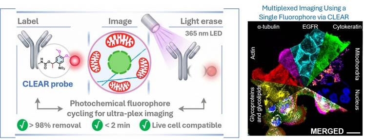
In simple terms, CLEAR turns a biological sample into a reusable imaging surface. Instead of trying to fit many fluorescent labels into one crowded image, the platform lets scientists capture one layer of information, erase it cleanly, and then capture the next layer. The result is a detailed protein atlas of the same cell or tissue section, built step by step with high spatial precision.
The platform was developed by a team led by Prof. Sarit S. Agasti at JNCASR. The team designed and synthesised the CLEAR probes, created the imaging workflow and validated the system across different biological settings. Researchers from the Indian Institute of Science also contributed to demonstrating the platform in more complex biological contexts, including immune cell systems.
For cancer research, the potential is substantial. Tumours are highly complex environments. Cancer cells interact with immune cells, blood vessels, stromal cells and signalling molecules in ways that influence disease progression and treatment response. A platform that can map many proteins in the same tissue section can help researchers understand tumour architecture with far greater depth. This could support better classification of tumours, sharper identification of disease markers and eventually more informed therapeutic decisions.
The relevance to neurological disorders is equally important. The brain is built on dense cellular networks where protein location and organisation carry major biological meaning. Many neurobiological conditions involve subtle changes in cellular signalling, synaptic structure, inflammation and protein distribution. CLEAR’s ability to preserve spatial context while expanding protein visibility can help researchers study these changes with greater clarity.
Another promising area is immunology. The published study highlights the use of CLEAR for studying immunological synapse formation, where immune cells reorganise their internal structures and signalling machinery while interacting with target cells. Such processes are dynamic and complex, requiring high-dimensional imaging methods that can capture multiple molecular events in the same cellular setting.
The larger meaning of the breakthrough is that India is contributing to the global rise of spatial proteomics. Modern medicine is moving beyond isolated biomarker testing towards a fuller understanding of how molecules are arranged inside tissues. This is essential for precision medicine, where treatment decisions may increasingly depend on detailed molecular signatures rather than broad disease labels. CLEAR fits into this direction by offering a faster, gentler and more scalable route to high-content protein imaging.
At the same time, the technology should be understood as a research-platform breakthrough rather than an immediate hospital diagnostic product. Its clinical impact will depend on further validation, standardisation, integration with pathology workflows and adoption by biomedical laboratories. But the foundation is significant: a method that can repeatedly image proteins in the same sample with high efficiency, high resolution and sample compatibility.
The achievement also reflects the growing strength of Indian institutions in advanced chemical biology, microscopy and biomedical technology. JNCASR and IISc are not merely participating in global science; they are helping define new methods that can be used internationally. The publication of the work in Chemical Science adds peer-reviewed weight to the development and places the research within the global conversation on next-generation imaging.
CLEAR’s greatest value may lie in the way it expands the researcher’s view of life at the microscopic level. A cell is not a static object. It is a crowded, active, highly organised molecular city. Disease changes that city’s traffic, architecture and communication signals. With CLEAR, scientists gain a sharper instrument to observe those changes layer by layer. For cancer biology, neurological research, immunology and precision medicine, that deeper view could become a major step towards earlier detection, better understanding and more targeted treatment strategies.
Publication Link: 10.1039/D5SC08599C
You may also like
-
Indian Researchers Study RAD51 DNA-Repair Gene Variant in Prostate Cancer
-
TDB Backs Multi Nano Sense With ₹37.51 Crore to Build Indigenous Multi-Gas Sensor Platform
-
IISER Berhampur and NIT Rourkela Researchers Explore Cotarnine Derivatives for Wound Healing and Biomedical Applications
-
IISER Tirupati Develops pH-Responsive DNA Nanostructures for Controlled Drug Delivery
-
India and France Expand Space Cooperation Into Human Spaceflight, TRISHNA and Start-up Ecosystem
